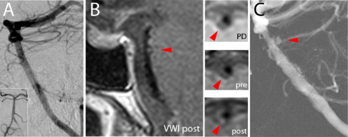
Figure 2.
A-C: (A) Initial lateral projection digital subtraction angiogram of the basilar artery was unrevealing, presumably due to thrombosis of the aneurysm at the time of imaging. Inset shows an anteroposterior projection. (B) Vessel wall MR imaging showed a focal outpouching with punctate enhancement along the basilar artery posterolateral wall (arrowhead) on postcontrast imaging. Insets show axial proton density, axial T1-weighted precontrast, and axial T1-weighted postcontrast images through the basilar artery outpouching with punctate enhancement. (C) Repeat lateral projection digital subtraction angiogram of the basilar artery showed a 2 mm perforator aneurysm.

