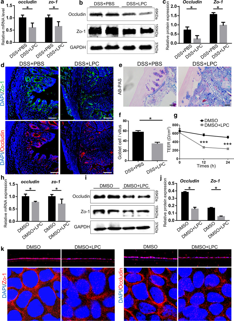
Fig. 9.
LPC impaired intestinal barrier function in vitro and in vivo. a Relative mRNA and b, c Protein expression of occludin and ZO-1 in colon tissues. GAPDH was used as a loading control. d The typical immunofluorescent images of colon tissues stained with ZO-1 (green), and occludin (red) in each group (Scale bar, 100 μm). e, f The typical images of colon tissue stained with AB/PAS and the number of goblet cells from two groups (Scale bar, 100 μm). g TEER of Caco-2 cells treated or not treated with LPC. (H) The mRNA level of ZO-1 and occludin in caco-2 cells treated with LPC of two groups. i, j The protein levels of ZO-1 and occludin in caco-2 cells treated with LPC of two groups. k The immunofluorescent images of Caco-2 cells with ZO-1 and occludin in each group (Z-axis and XY-axis). Data were shown as mean ± SEM (n = 6 per group). The data come from three independent experiments. In all panels: *p < 0.05. (AB/PAS alcian blue/periodic acid Schiff, TEER transepithelial/transendothelial electrical resistance)

