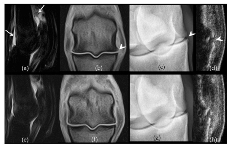
Figure 3.

Comparative imaging findings observed on week 12 on the front metacarpophalangeal joints of horse 7. The left fore fetlock (a–d) received placebo treatment while the right fore fetlock (e–h) received bone-marrow derived mesenchymal stem cells (BM-MSCs) on week 3. On the left fetlock there was grade 3 (white arrows) synovial effusion (a) and periarticular osteophytes (b) (arrow heads) on Short Tau Inversion Recovery sagittal magnetic resonance imaging (MRI) images (a) and T1-weighed dorsal MRI images (b) respectively while grade 2 synovial effusion was observed on the right fetlock (e–f). Similarly, there are grade 3 (arrow head) osteophytes on the dorsomedial-palmarolateral 35° oblique radiographic view from the left fore fetlock (c) compared to a grade 1 on the right (g). The ultrasound osteophyte score was 3 on the left (d) (arrow head) and 2 on the right fetlock (h), on the longitudinal sections made on the dorso-lateral aspect of both joints.
