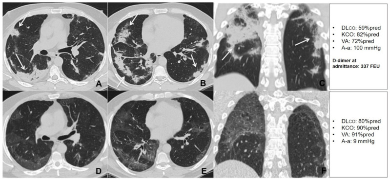
Figure 7.
Unenhanced thin section axial (A,B) and coronal (C) CT images of the lungs obtained the 10th day post-admission in a 51-year-old man with no previous exposure to cigarette smoke, no cormorbidities, that was admitted to the high dependency respiratory care unit (HDRU) with severe respiratory failure (PaO2/FiO2 182 mmHg) and treated with continuous positive airway pressure (CPAP) for 10 days. The images show peripheral consolidations (arrows) in both lungs, with predilection for posterior areas. Unenhanced thin section axial (D,E) and coronal (F) CT images of the same patient 6 weeks post-discharge show bilateral peripheral GGO with resolution of previous consolidations. The patient had a low D-dimer at admission and showed a significant improvement in his DLco during the recovery period.

