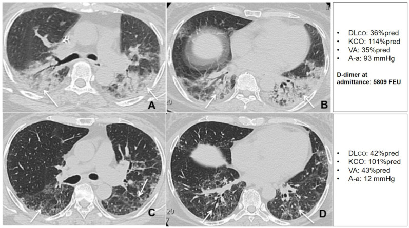
Figure 8.
Unenhanced thin section axial (A,B) CT images of the lungs obtained the 12th day post-admission in a 57-year-old woman with arterial hypertension and no previous exposure to cigarette smoke, that was admitted to the HDRU with a PaO2/FiO2 of 210 mmHg, rapidly worsened her respiratory status and needed invasive mechanical support for 8 days. The images show peripheral posterior GGO and consolidations in both lungs, with predilection for posterior areas (arrows). Unenhanced axial (C,D) CT images of the same patient 6 weeks post-discharge show residual interlobular septal thickening with intralobular lines (arrows) with resolution of previous GGO and consolidation. The patient had a high D-dimer at admission and showed very limited changes in DLco during the recovery period.

