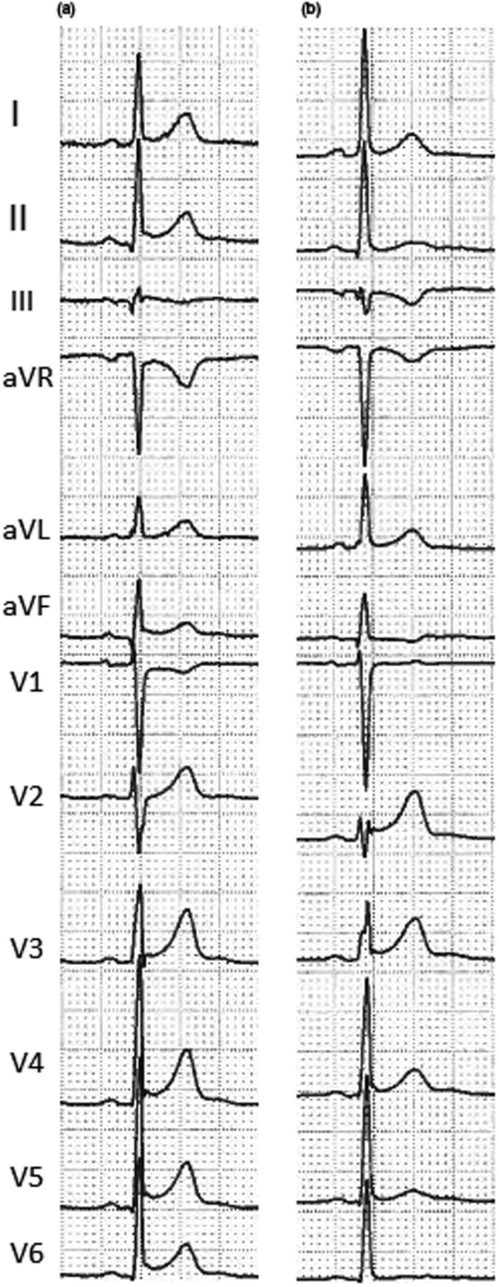
Figure 2.

Electrocardiogram performed at admission (a) and 3 days later (b) in a 30‐year‐old patient with perimyocarditis. Comparison between these two ECGs shows the following: disappearance of ST elevation with pericarditis pattern in inferior and lateral leads, disappearance of ST depression in aVR lead, and onset of T‐wave inversion in III e aVF leads
