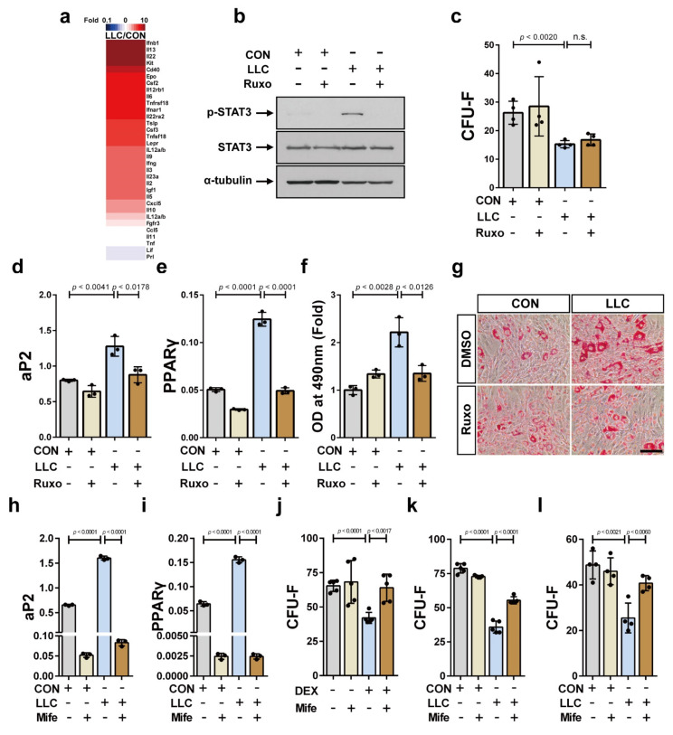
Figure 4.
The activation of JAK/STAT and glucocorticoid signaling are associated with the depletion and the unbalanced differentiation capacity of bone marrow MSCs in LLC-cancer cachexia, respectively. (a) Protein content in the serum of LLC-bearing mice at 25 days postinoculation of LLC cells (LLC) and the control mice at 25 days post-sham-operation (CON) was determined using protein antibody arrays. Proteins related to JAK/STAT signaling activity were analyzed. Thirty-one proteins in the serum of LLC-bearing mice that showed a difference of at least 1.3 times compared to the value in the control (p value < 0.05) were presented. (b) Western blot analysis was performed using ST2 cells that had been treated with serum derived from LLC-bearing (LLC) or the control mice (CON) for 24 h after pretreatment with ruxolitinib (+ Ruxo) or solvent (- Ruxo) for 30 min. Uncropped Western blot images are available in Figure S2. (c) CFU-F assay was performed using ST2 cells that had been treated with serum derived from LLC-bearing mice (LLC) or the control mice (CON) for three days in addition of ruxolitinib (+ Ruxo) or solvent (- Ruxo). The expression levels of aP2 mRNA (d) and PPARγ mRNA (e) were analyzed after the adipogenic differentiation of ST2 cells using differentiation inducing media supplemented with serum derived from LLC-bearing mice (LLC) or the control mice (CON) in addition of ruxolitinib (+ Ruxo) or solvent (- Ruxo). (f,g) The adipogenic differentiation of ST2 cells was also confirmed with Oil Red O staining (Scale bar, 100 μm). The expression levels of aP2 mRNA (h) and PPARγ mRNA (i) were analyzed after the adipogenic differentiation of ST2 cells using differentiation inducing media supplemented with serum derived from LLC-bearing mice (LLC) or the control mice (CON) treated with mifepristone (+ Mife) or solvent (- Mife). (j) CFU-F assay was performed using ST2 cells that had been treated with dexamethasone (DEX, 1 μM) for 3 days with the addition of mifepristone (+ Mife) or solvent (- Mife). CFU-F assay was performed using ST2 cells (k) or human bone marrow MSCs (l) that had been treated with the culture media supplemented with serum derived from LLC-bearing mice (LLC) or the control mice (CON) for three days with the addition of mifepristone (+ Mife) or solvent (- Mife). Data are presented as the mean ± SD (p values were obtained by t-tests. n.s.; not significant).

