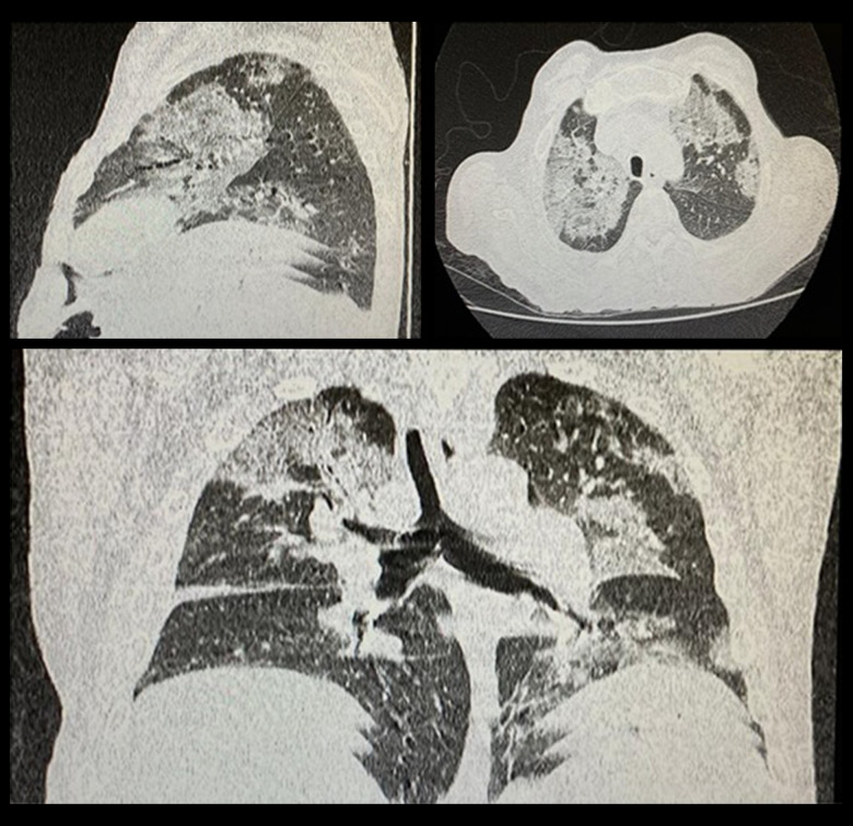
Figure 1.
Clockwise, from left to right: sagittal, axial, and coronal section. They demonstrate multiple ground-glass opacities, predominantly perihilar, in the middle and upper third of the pulmonary fields, associated with thickening of the inter- and intralobular septa, which forma mosaic paving pattern.

