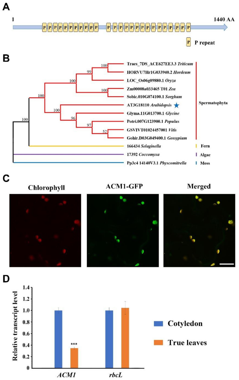
Figure 3.
Sequence analysis and subcellular localization of ACM1. (A) Schematic diagram of ACM1 protein with 26 PPR domains (P). (B) Phylogenetic tree of ACM1 and other 12 PPR family members from Figure S1. Maximum likelihood (ML) tree was inferred with RAxML using the PROTGAMMALGF model. The numbers on the branches refer to the bootstrap values (%) for 1000 replications. Complete deletion was adopted for the treatment of gaps and missing data. (C) Localization of the ACM1 protein within the chloroplast using the GFP assay by transient transformation into tobacco leaf epidermal cells. Scale bar: 10 μm. (D) Quantitative real-time PCR to detect the expression profile of ACM1 transcript between cotyledons and true leaves in WT. RNA was extracted from 7-day-old cotyledons and 14-day-old true leaves from WT, and then reverse-transcribed. *** P < 0.001, by Student’s t-test.

