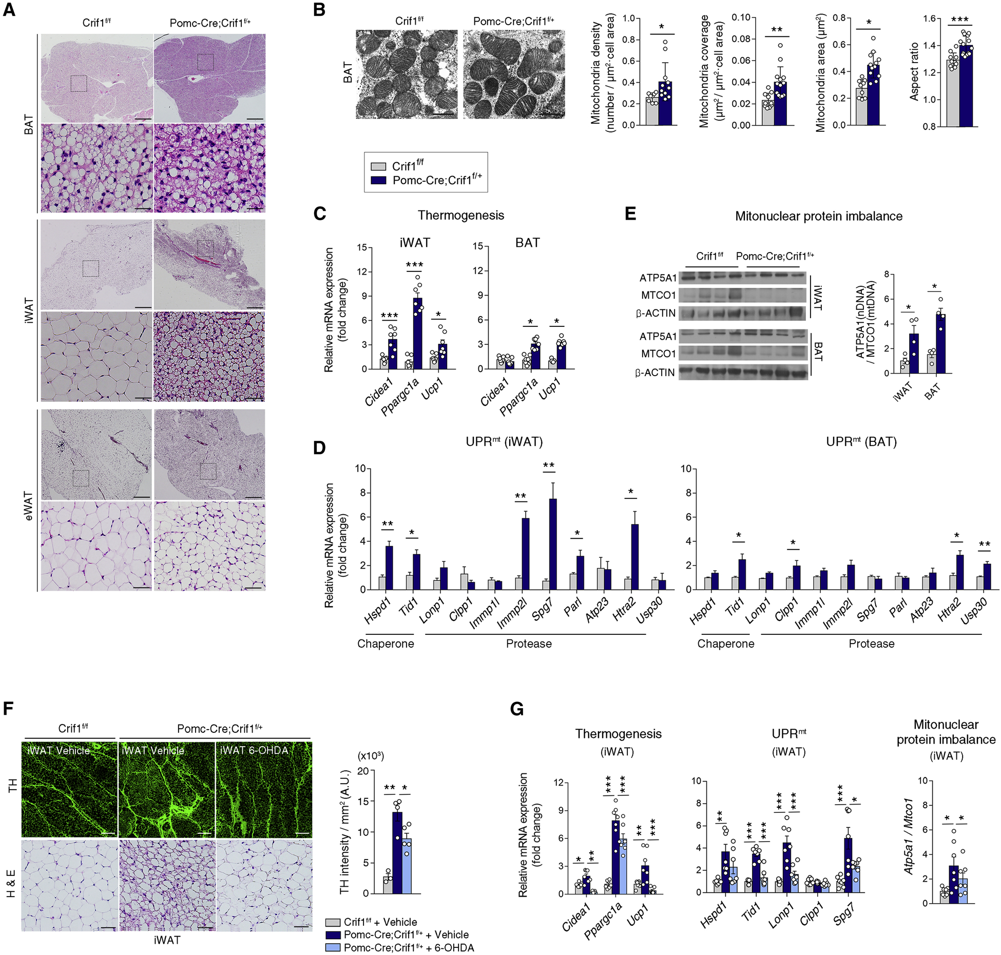
Figure 3. Mild mitoribosomal stress in POMC neurons activates a thermogenic program and UPRmt in adipose tissues.

(A) Histology of brown adipose tissue (BAT), inguinal white adipose tissue (iWAT) and epididymal white adipose tissue (eWAT) in Crif1f/f and Pomc-Cre; Crif1f/+ males. Scale bars, 500 μm (upper) and 50 μm (lower).
(B) Electron microscopic examination of the BAT from 8 week-old Crif1f/f and Pomc-Cre; Crif1f/+ males (n = 8–13 cells). Scale bars, 0.5 μm.
(C) Thermogenesis-related gene expression in the iWAT and BAT at 8 weeks (n = 7).
(D) Increased mitochondrial chaperone and protease expression in the iWAT and BAT of Pomc-Cre; Crif1f/+ males, indicating a mitochondrial unfolded protein response (UPRmt) (n = 4).
(E) Mitonuclear protein imbalance between nuclear DNA (nDNA)-encoded ETC protein (ATP5A1) versus mitochondrial DNA (mtDNA)-encoded protein (MTCO1) in the iWAT and BAT (n = 4).
(F, G) Reversal of iWAT browning, increased sympathetic innervation and thermogenic gene expression, the UPRmt and the mitonuclear protein imbalance in Pomc-Cre; Crif1f/+ males following an intra-iWAT injection of neurotoxin 6-hydroxydopamine (OHDA). Tyrosine hydroxylase (TH) staining of sympathetic innervation in the iWAT is shown. Scale bars, 200 μm (upper) and 50 μm (lower) (n = 6–7).
Results are presented as a mean ± SEM. *p < 0.05, **p < 0.01, and ***p < 0.001 between indicated groups. See also Figure S3.
