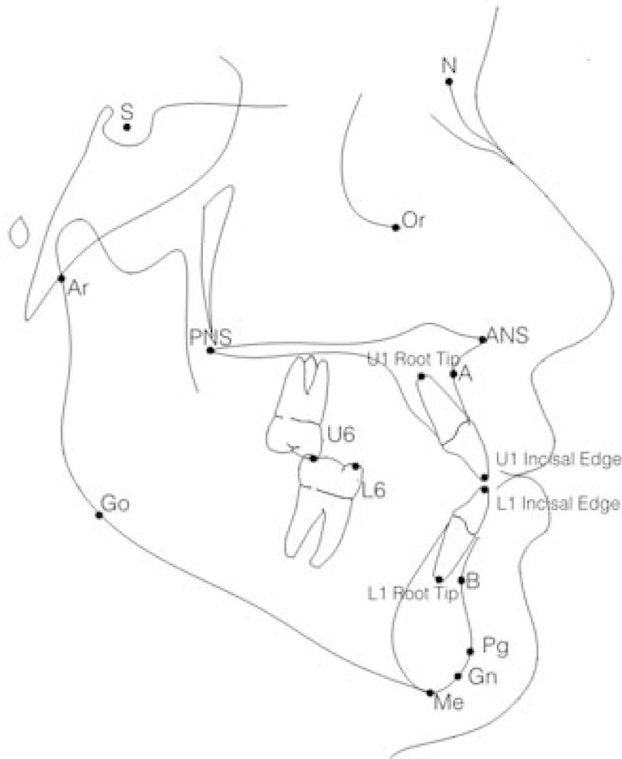
Figure 1.

Cephalometric landmarks identified on pre- and post-treatment lateral cephalograms: sella (S), nasion (N), anterior nasal spine (ANS), posterior nasal spine (PNS), pogonion (Pg), gnathion (Gn), menton (Me), anatomic gonion (Go), articulare (Ar), A-point (A), B-point (B), incisal edge of the maxillary incisor (U1 incisal edge), root tip of the maxillary incisor (U1 root tip), incisal edge of the mandibular incisor (L1 incisal edge), root tip of the mandibular incisor (L1 root tip), mesiobuccal cusp tip of the maxillary first molar (U6), mesiobuccal cusp tip of the mandibular first molar (L6).
