
FIG 1.
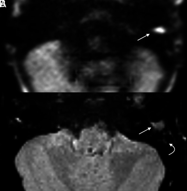
Cholesteatoma with positive findings on both HASTE and RESOLVE in a 4-year-old girl with delayed speech. Both axial HASTE (A) and RESOLVE (B) demonstrate a region of restricted diffusion in the left external auditory canal (arrow). Note greater signal hyperintensity of HASTE compared with the RESOLVE image. Adjacent opacified mastoid air cells demonstrate relatively faint signal (curved arrow).