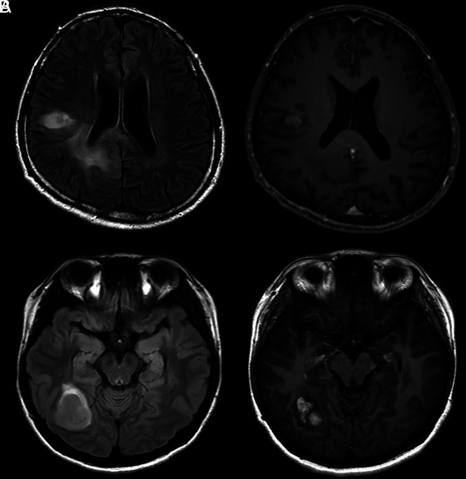
FIG 2.
Representative cases of IDH wild-type lower-grade gliomas with (A) and without (B) molecular features of GBM. A, Initial MR imaging of a 49-year-old man with IDH wild-type lower-grade glioma with molecular features of GBM, WHO grade III. MRIs reveal infiltrative T2-hyperintense tumor in the right frontal and parietal lobes extending into the corpus callosum with cortical involvement (left). Focal contrast enhancement is noted in the right frontal lobe (right). B, Initial MRIs of a 26-year-old woman without molecular features of GBM, WHO grade III. MRIs reveal a relatively well-defined enhancing mass with peritumoral edema in the right temporo-occipital lobe without cortical involvement.

