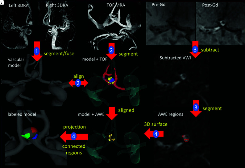
FIG 1.
Illustration of methodology with the anterior communicating artery aneurysm. Step 1: model construction from 3DRAs (A–C). Step 2: rough segmentation of TOF-MRA and alignment with the vascular model (D and E). Step 3: pre- and post-Gd MR image subtraction and AWE region segmentation (F–I). Step 4: 3D-AWE region isosurface extraction, alignment, projection to the vascular model, and splitting into connected components (J and K). Models constructed from CTAs follow a similar approach, while models from TOF-MRA are automatically aligned to the Gd MR imaging series. VWI indicates vessel wall imaging.

