Fig. 4.
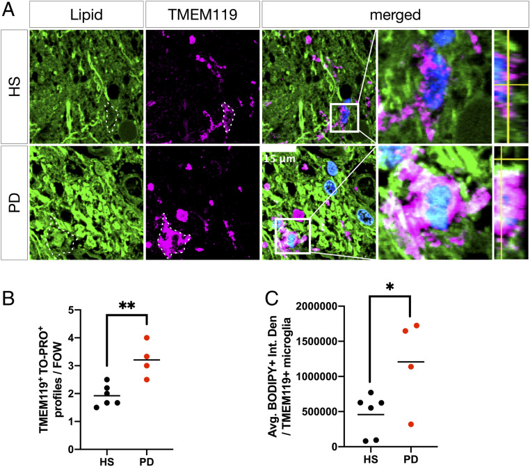
Microglia accumulate neutral lipids in PD SN. (A) Representative micrographs of BODIPY (green) and TMEM119 (magenta) colabeled microglia in HS SN and PD SN. Boxed insets are shown in higher magnification with orthogonal views on the right. Dashed lines indicate TMEM119+ microglial cell outlines. Nuclei (TO-PRO; blue) are shown in merged views (scale bar: 15 µm). (B) Average number of TMEM119+/TO-PRO+ microglia per randomly chosen field of view. **P < 0.01, n = 4 to 6 subject averages per group, n = 4 to 6 fields of view per subject. (C) Quantification of BODIPY+ neutral lipid (integrated density) associated with TMEM119+/TO-PRO+ microglial cell profiles. *P < 0.05, n = 4 to 6 subject averages per group, n = 5 to 10 microglial cell profiles per subject.

