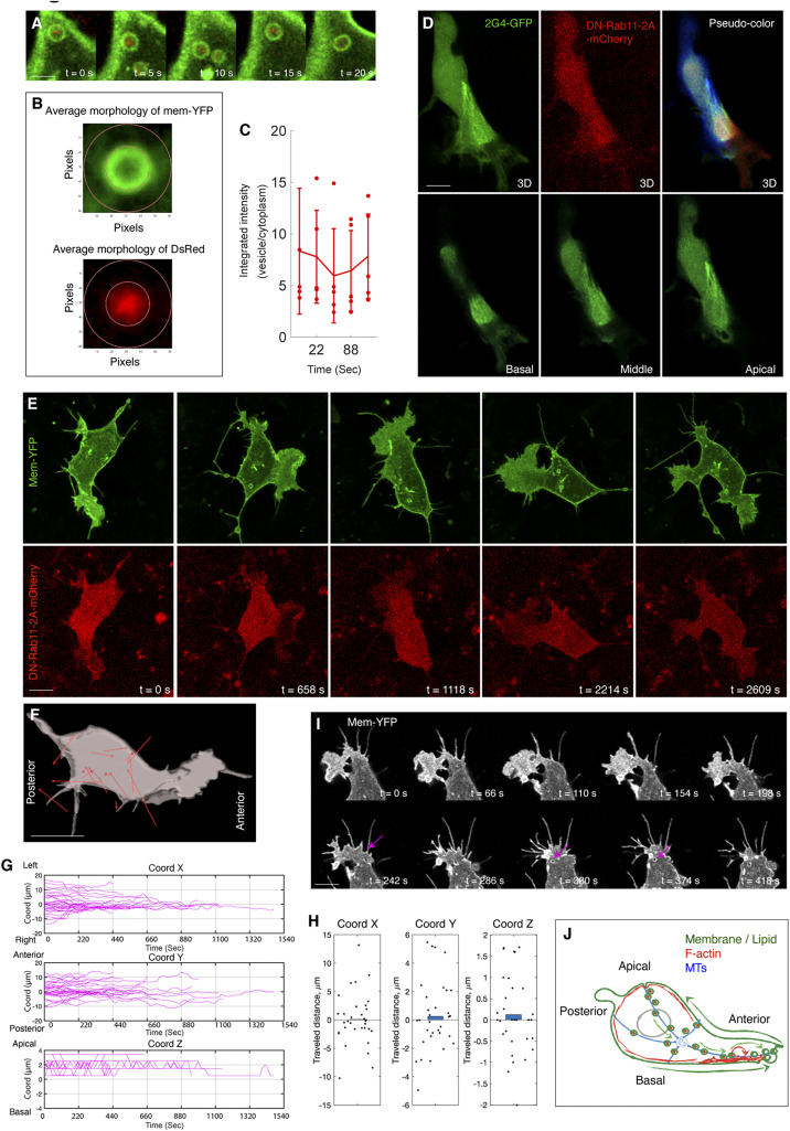
Fig. 7.
Blocking Rab11 activity interferes vesicle flow and lamellipodial maintenance. (A–C) Rab11 is colocalized with vesicles. (A) Live imaging to show Rab11-DsRed labeled puncta (red) is enriched in the vesicles. B illustrates the principle of colocalization analysis. The inner ring is drawn along the border of Membrane-YFP (green). The red pixels in the inner ring and in the region between the inner and outside rings are measured. The ratio between them is plotted (C); if the ratio is more than one, it means more Rab11 inside the vesicles than in the cytoplasm. (Scale bar in A: 1 µm.) (D) MT organization in a DN-Rab11-2A-mCherry–positive cell. Three individual slices of the cell (from basal to apical side) are presented. In the 3D image (Upper Right), each individual slice is pseudocolored. Similar to the untreated cells in SI Appendix, Fig. S2B. (E) Live imaging of a DN-Rab11-2A-mCherry–expressing cell shows lamellipodial extension in multiple directions. (F) Net displacement vectors of the cell in E showing that vesicle movement is not directional. (Scale bars in D–F: 2 µm.) (G) Trajectory analysis of the vesicles in the DN-Rab11–positive cells. Along the y axis, the lines display both upward and downward shifts, the evidence of vesicle migration to either end of cells. n = 35 vesicles, n = 3 cells. (H) Distance analysis shows these vesicles move in shorter distance than the normal ones in Fig. 2G, n = 35 vesicles, n = 3 cells (rank sum test, P < 0.001, x: P = 0.905; y: P = 0.543; z: P = 0.433). (I) Live imaging of noncanonical macropinocytosis in a DN-Rab11–expressing cell. Similar to both untreated (Fig. 4B) and nocodazole treated cells (Fig. 6J), tail retraction produces small vesicles (arrows). (Scale bar: 3 µm.) (J) Schematic summary of the membrane and cytoskeletal processes in controlling cell polarization and locomotion.

