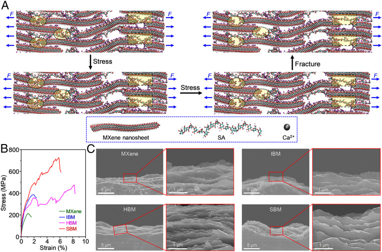
Fig. 4.
Fracture mechanism of the SBM sheets. (A) Model snapshots for the SBM sheets during simulative tensile stretching showing not only the breakage of hydrogen bonding and stretching of coiled SA molecular chains (yellow-shaded rectangular region) but also the breakage of ionic bonding (yellow-shaded elliptical region). (B) Simulated stress–strain curves for the MXene, IBM, HBM, and SBM sheets. (C) Inclined-view SEM images of the fracture surface of the MXene, IBM, HBM, and SBM sheets, indicating curling of the platelet edges for the IBM, HBM, and SBM sheets but not for the MXene sheets.

