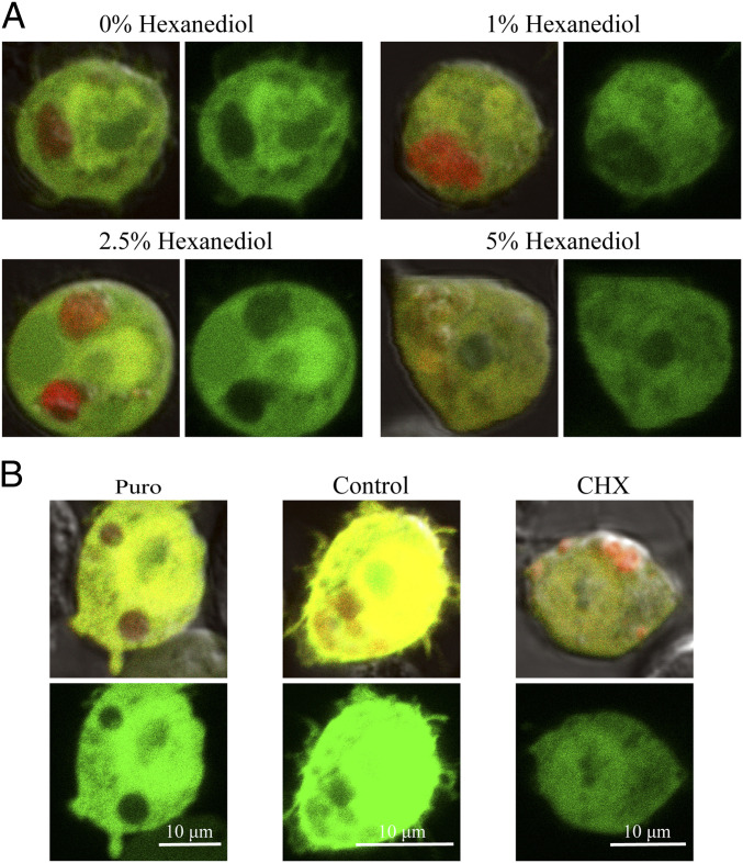
Fig. 4.
AfrLEA6 condensates behave like stress granules in vivo. Colocalization of GFP with or exclusion from AfrLEA6 condensates is visualized by comparing images that overlay red (mCherry) and green (GFP) fluorescence with images showing only the green fluorescence signal (Right). (A) Cells expressing AfrLEA6 tagged with mCherry concurrently with GFP were incubated for 1 h with 0 to 5% (wt/vol) of 1,6-hexanediol in DPBS. (Images to the Left show overlay of red and green fluorescence and images to the Right show green fluorescence only.) (B) Cells expressing AfrLEA6-mCherry were subjected to either puromycin (25 µg/mL) or CHX (100 µg/mL) for 2 to 3 h. (Top images shows overlay of red and green fluorescence and Bottom image shows green fluorescence only.)

