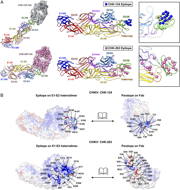
Fig. 4.
Epitopes bound by CHK-124 Fab and CHK-263 Fab. (A) Binding mode of CHK-124 Fab (Top) and CHK-263 Fab (Bottom) to the ectodomains of an E1-E2 heterodimer. Epitopes of CHK-124 and CHK-263 are shown as blue and pink spheres, respectively, on an E1-E2 heterodimer (Middle and Right). (Right) Zoom-in view of the epitope with strands labeled. (B) Open book representation showing the electrostatic potential of the interacting interface between the epitope and paratope of CHIKV:CHK-124 (Top) and CHIKV:CHK-263 Fab (Bottom) structures. Positive, negative, and neutral charged residues are colored in blue, red, and white, respectively. Cyan dashed lines indicate the boundaries of the antibody heavy and light chains in the paratope and also its corresponding binding epitope. Residues at the interacting interface are indicated.

