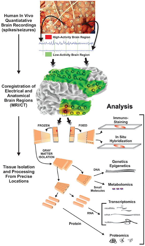
Figure 1. Human epilepsy systems biology workflow.

Patients who undergo long-term intracranial EEG monitoring followed by tissue removal to treat their epilepsy provide a unique opportunity to discover what is different about epileptic brain areas compared to non-epileptic brain areas. Precisely localized brain regions with high or low epileptic activities (spiking, non-spiking plus or minus seizures) quantified by a variety of algorithms are co-registered to MRI and CAT scans which aligns electrode locations with brain structures. Tissue blocks (1 cm3) from resected tissues at each electrode location are subdivided so that half is fixed for histology and the other half frozen immediately for molecular analyses. Using this approach, immunostaining and in situ hybridization are performed on the fixed tissue, whereas genomics, proteomics, and metabolomics are performed on alternating strips of even amounts of layers I-VI from the gray matter. In this way the molecular measures can be directly linked to histology, brain imaging, quantitative electrophysiology, and patient clinical information.
