Figure 6.

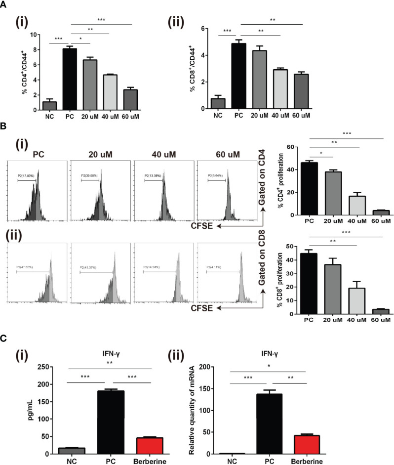
The effects of berberine on CD4+ and CD8+ T cell apoptosis, activation, and proliferation in vitro. (A) T cell activation assay. The percentages of (i) CD4+CD44+ and (ii) CD8+CD44+ T cells were determined by flow cytometry. (B) T cell proliferation assay. T cells from naïve C57BL/6 mice were labeled with CFSE and then co-stimulated with anti-CD3/CD28 Abs in the absence or presence of berberine. After 3 days of co-culture, (i) CD4+ and (ii) CD8+ T cell division was determined by flow cytometry. (C) (i) Supernatant levels of IFN-γ measured by ELISA, and (ii) mRNA expression of IFN-γ in T cells measured by qPCR. *p < 0.05, **p < 0.01, ***p < 0.001 compared to the (Positive Control) PC group.
