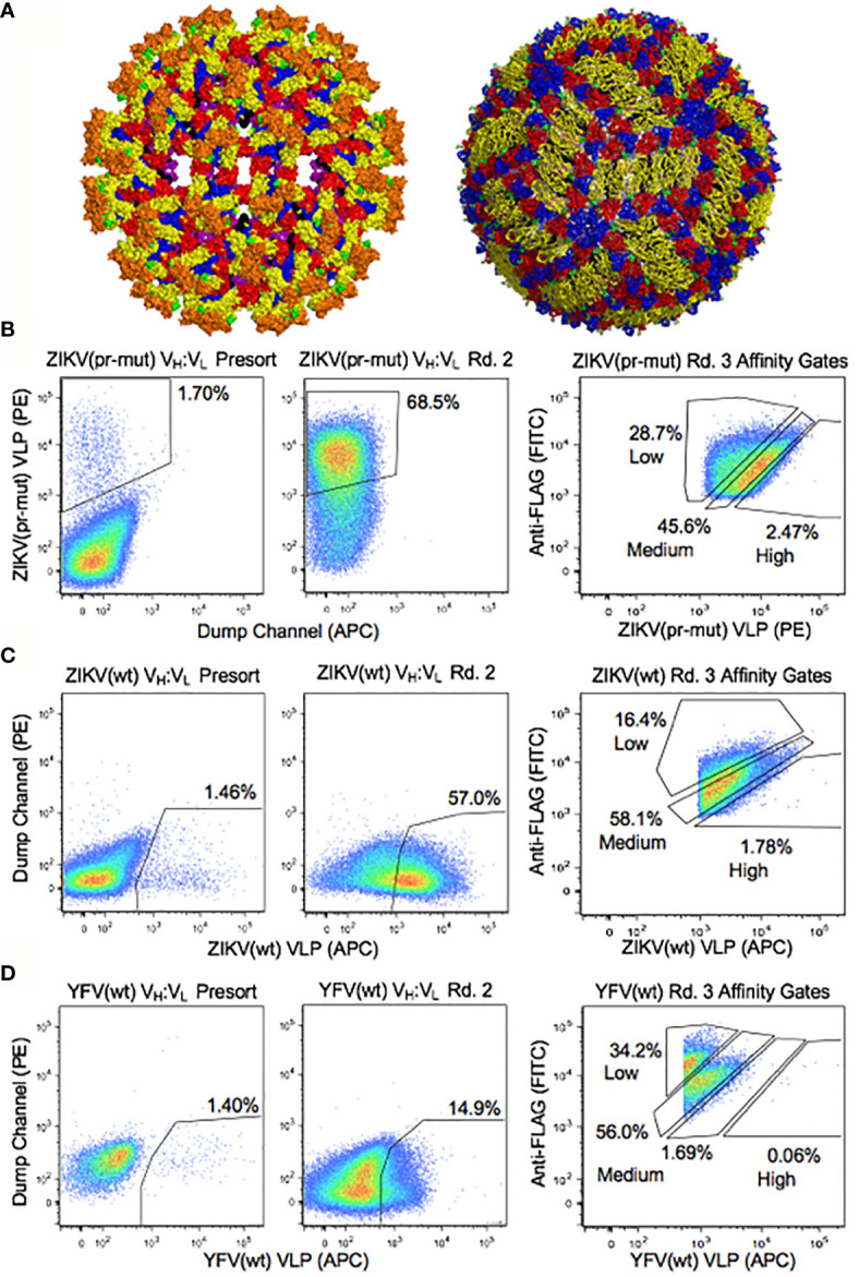
Figure 2.

Functional analyses of yeast display repertoires for binding to flavivirus antigens. (A) Surface representation of ZIKV envelope proteins on the immature (left) (57) and mature (right) (58) forms of the native virus. Colors indicate E protein domain I (red), domain II (yellow) and domain III (blue), fusion loop of domain II (green), transmembrane region of E protein (purple), M protein (black), and pr domain (Orange). Functional screening was performed with virus-like particles ( Figure S1 ), and the solved structures of the native virus are shown here for reference. (B–D) Representative FACS analysis of yeast display repertoires from B cells of ZIKV disease convalescent patients screened for binding to (B) ZIKV (pr-mut), (C) ZIKV (wt), and (D) YFV (wt) VLPs. FACS affinity gates were used to bin repertoires by antigen affinity.
