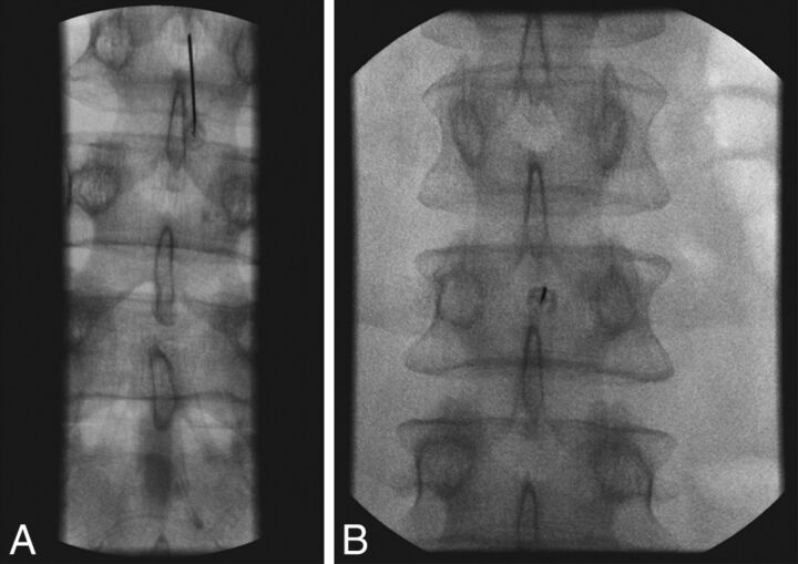
Fig 8.
Fluoroscopic spot films obtained during LP, showing parallax artifacts. A, Note that on this image, the needle is not centered and the tip appears to abut the inferior spinous process. B, The same procedure with no needle adjustment, but now the tip is centered in the image. CSF flow was normal.

