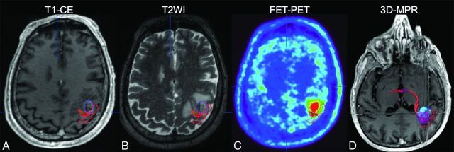
Fig 1.
A 72-year-old male patient with contralateral upper limb weakness as depicted by an overlap between his carcinoma-metastasis on T1-CE (purple in A, B, and D) and FET-PET (light blue in C and D) and the hand-motor representation (red in A–D). 3D multiplanar reconstruction image with integration of functional, metabolic, and anatomic data is shown in (D).

