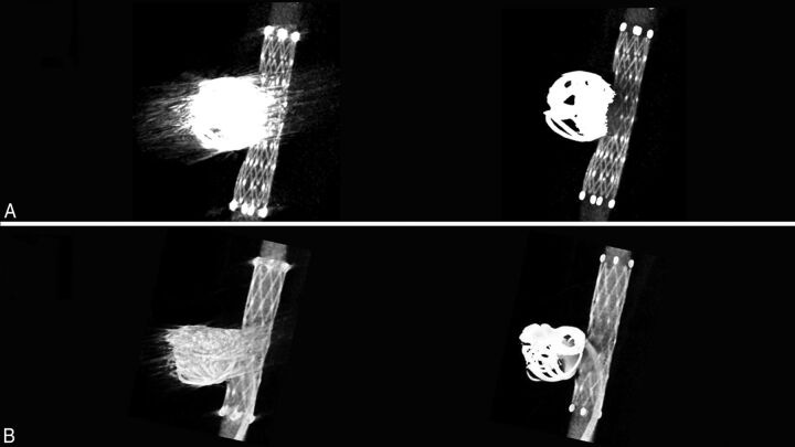
Fig 2.
Thin-section MIP images of the aneurysms treated with stent-assisted coil embolization. A, Comparison of the pre- (left) and post-MAR (right) images of an aneurysm treated with a combination of bare platinum coils and a Neuroform stent (4 × 20 mm) reveals improved visibility of the stent structures near the coil mass after MAR processing. B, Another aneurysm that was treated with coil embolization by using a Neuroform stent. Note the improved visibility of the microcatheter inserted in the aneurysm and contrast filling in the aneurysm after the MAR processing (right).

