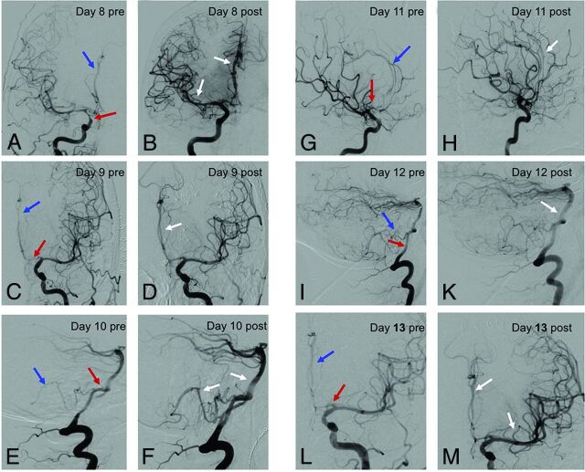
Fig 1.
Repeat angiograms in refractory cerebral vasospasms. Repeat angiograms (red arrows: proximal spastic vessels; blue arrows: distal spastic vessels) in a 41-year-old woman with a ruptured anterior communicating artery aneurysm initially treated with coil embolization. On day 8 after the ictus, the patient presented with hemiparesis on the left side. A, ICA angiogram, anteroposterior view, shows vasospasm of the right-sided M1 and A1/A2 segments. B, ICA angiogram of the same patient after nimodipine infusion demonstrates reduced vasospasm of the M1 and A1/A2 segments, with improved perfusion of both the proximal and distal arteries. C–M, Corresponding angiograms of the anterior circulation (C, D, G, H, L, M) and posterior circulation (E, F, I, K) in the same patient during the CVS period from day 9 to day 13. Images on the right side show the effect after IAN. Note the effect of nimodipine infusion on the vessel diameter (white arrows).

