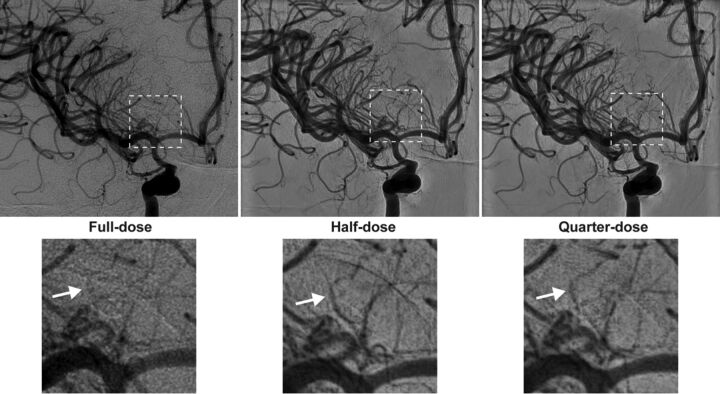
Fig 3.
Diagnostic angiography was performed to assess the source of bleeding in a 43-year-old man who presented with diffuse SAH. During the examination, angiograms after contrast administration into the right ICA were obtained on IPDRT platform by using the “full-dose” protocol (left), which is identical to the reference platform in terms of hardware and software settings, “half-dose” protocol (middle), and “quarter-dose” protocol (right). Magnified views of the dashed area highlight improved visualization of small perforators (white arrows) with the “half-dose” and “quarter-dose” protocols (lower panels).

