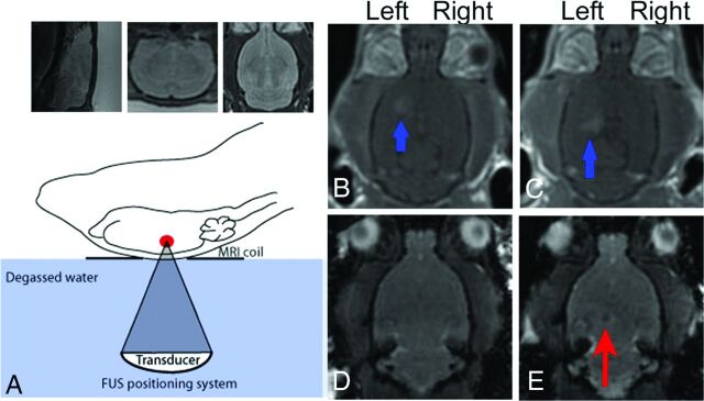
Fig 3.
Application of MRgFUS for the delivery of iron-labeled neural stem cells. Schematic of the FUS apparatus (lower left). Sagittal, coronal, and axial T2-weighted images (A) are used to identify the intended targets of neural stem cell delivery in the left hippocampus and left striatum. T1-weighted, postcontrast images after local FUS sonication (B and C) demonstrate enhancement in the striatum and hippocampus (blue arrows) compatible with enhanced BBB permeability. Fast gradient-echo sequences are obtained before (D) and after (E) sonication, localizing a focus of hypointense signal (red arrow) confirming delivery of iron-labeled neural stem cells. Reprinted with permission from Burgess et al.40

