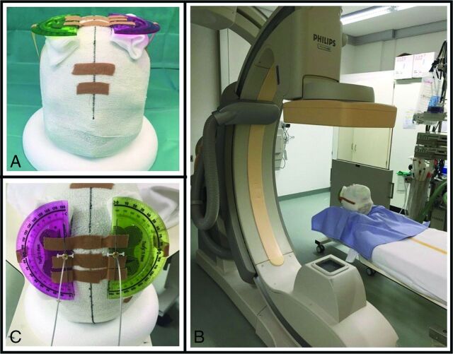
Fig 1.
Two directional leads inserted in a 3D-printed plaster head filled with gelatin. At the entry site of the electrodes, a marker was attached in the same orientation as the x-ray marker at the electrode tip. The electrodes were guided through protractors (A,C), which allows for determining the rotation of each electrode between 0° and 360°(C, both oriented at 0°). Figure 1B shows the setup in the 3D rotational fluoroscopy.

