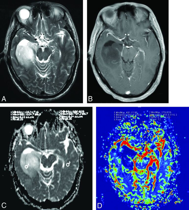
Fig 2.

A 50-year-old man with an anaplastic astrocytoma with an IDH mutation. A, Axial T2WI demonstrates heterogeneous high signal intensity with sharp borders on the right temporal lobe. B, Contrast-enhanced axial T1-weighted image demonstrates a nonenhancing lesion in the right temporal region. C, A corresponding ADC map shows the tumor with an increased ADC value (ADCmin = 1.456 × 10⁻³mm2/s, rADC = 2.51). D, Correlative color CBV image shows relatively low perfusion with the calculated rCBVmax of 1.86.
