Fig 1.
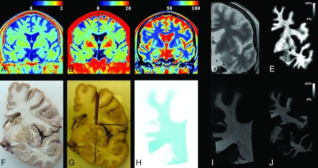
The process used for myelin evaluation on a male subject, 69 years of age, acquired at a temperature of 10°C. The MR imaging quantification sequence provided the R1, R2, and PD maps of coronal slices of the cadaver (A–C). A synthetic proton density–weighted image was created for registration purposes using the R1, R2, and PD maps as input, resampled to 0.1 mm/pixel (D, zoomed in). The R1 map was corrected for temperature and then used, with the original R2 and PD maps, to generate the myelin partial volume map with the same algorithm as used for living subjects (E). For the histologic images, the brain was extracted and cut into coronal slices (F). Slices were fixated by using formaldehyde and cut into smaller pieces after fixation (G). The separate pieces of brain slices were stained with Luxol fast blue and photographed (H). The optical density of the photographs was converted to an intensity scale. These images were also resampled to 0.1 mm/pixel (I). All pieces were registered to the synthetic proton density–weighted image (J), so that each pixel from the slice photographs was at the same place as the corresponding MR imaging pixel. Finally, the resolution of both MR images and photographs was down-sampled to the original MR imaging resolution of 0.7 mm/pixel.

