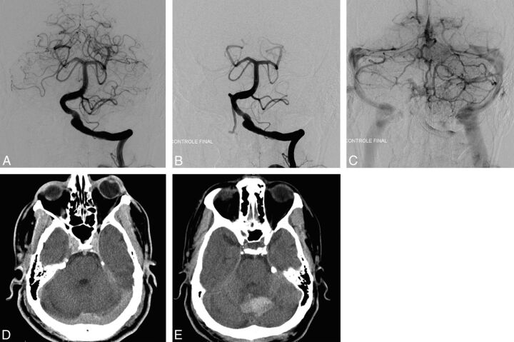
Fig 1.
A, Pretreatment angiography shows a left vertebral fusiform aneurysm. B and C, Postreatment angiography shows the placement and the permeability of the FD. D, Postreatment CT shows no postprocedural complications, mainly no intracranial bleeding. E, CT performed 7 days later for headache and aphasia reveals a vermian hematoma.

