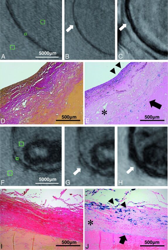
Fig 3.

Histopathologic images and MR imaging of both partially resected aneurysms are illustrated. There is an excellent correspondence between hypointense signal in TOF-MRA and SWI and iron deposition in the aneurysm wall in histopathologic sections (subject 3: A–E; subject 5: F–J). Green squares on TOF-MRA (without zoom interpolation) indicate ROIs in the aneurysm wall and surrounding structures (brain parenchyma and intraluminal thrombus) (A and F). Magnified images depict the hypointense aneurysm wall in TOF-MRA (B and G) and the strongly hypointense aneurysm wall in SWI (C and H). Aneurysm walls partially show the triple-layered microstructure (hyperintense layer in the middle) in both sequences (arrows). Histopathology shows aneurysm walls with Van Gieson elastic staining (original magnification ×50) (D and I) and iron deposition in inner smooth muscle (arrowheads) and adventitial layer (arrow) in Prussian blue staining (original magnification × 50) (E and J). Note the decellularized layer in the middle layer without iron deposition (asterisks).
