Figure.
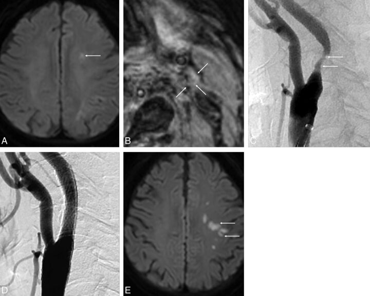
A 77-year-old man with a left acute embolic infarction due to severe stenosis of the left proximal internal carotid artery. A, Axial DWI at admission shows multiple areas of diffusion restriction in the left centrum semiovale (arrow). B, MPRAGE image shows isointensity of the left carotid plaque (arrows). This finding indicates a necrotic core without IPH. C, Lateral projection of left carotid angiography shows moderate stenosis in the proximal cervical portion of the left internal carotid artery (arrows). D, Carotid artery angiogram after protected CAS shows complete recanalization without residual stenosis. E, Axial DWI after CAS shows the new embolic lesions in the left frontal lobe (arrows).

