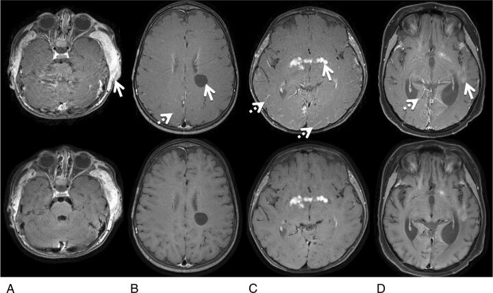
Fig 5.
Representative images with lesions from patients 2 (A), 8 (B), 15 (C), and 20 (D), comparing lesion conspicuity between Cartesian TSE (top) and spiral SE sections (bottom). A, The MR image shows extracranial soft-tissue enhancement related to postsurgical infection. B, The patient has a nonenhancing neural cyst. C, The patient has an enhancing hypothalamic glioma. D, The patient has scattered enhancing intracranial leukemia tumors. The solid arrows in the Cartesian images point to these lesions, and the dotted arrows point to noticeable bright-blood signals in the small vessels that are absent in spiral SE images.

