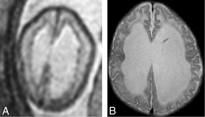
Fig 3.
An example of subependymal nodularity giving the false appearance of SEH (false-positive finding). Axial T2 single-shot fast spin-echo image from fetal MR imaging at 24 weeks' GA (A) demonstrates nodularity along the ependymal surfaces of the lateral ventricles, giving the appearance of SEH. However, postnatal MR imaging at 8 weeks of age (B) does not demonstrate any SEH.

