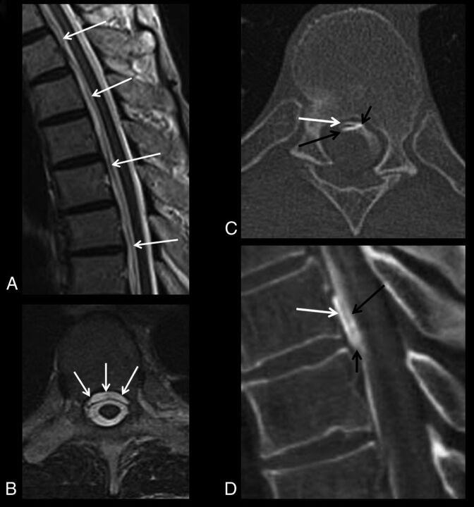
Fig 1.
Example of a patient with algorithm compliance and success. Initial spinal MR imaging demonstrates a ventral epidural fluid collection in the midthoracic spine on sagittal (white arrows, A) and axial (white arrows, B) T2-weighted images. The patient was appropriately triaged to dynamic CTM, in which a fast CSF leak is identified on initial dynamic axial (C) and sagittal reformatted (D) CT scans. C and D, Short black arrows indicate the CSF leak site; white arrows, ventral epidural contrast; and long black arrows, contrast in the ventral thecal sac.

