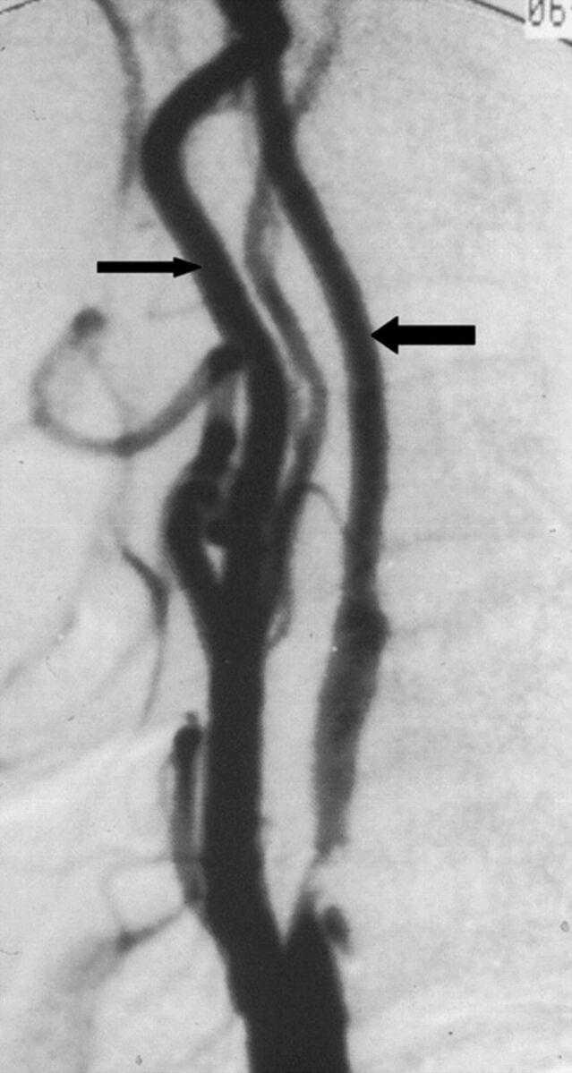
Fig 2.

A case with near-occlusion without full collapse, reprinted with permission from Fox et al.1 Lateral carotid angiogram shows a reduced ICA lumen distal to the stenosis (larger arrow); the diameter is slightly less than the ECA diameter (smaller arrow). The distal ICA lumen is normal-appearing (not threadlike).
