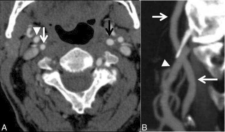
Fig 4.
A case with near-occlusion without full collapse. A, Axial CTA at the level of the distal extracranial ICA. B, Sagittal reformat of A. The distal right ICA (white arrow) is narrower than the contralateral ICA (black arrow) and similar to the right ECA (arrowhead) but otherwise is normal-appearing (not threadlike).

