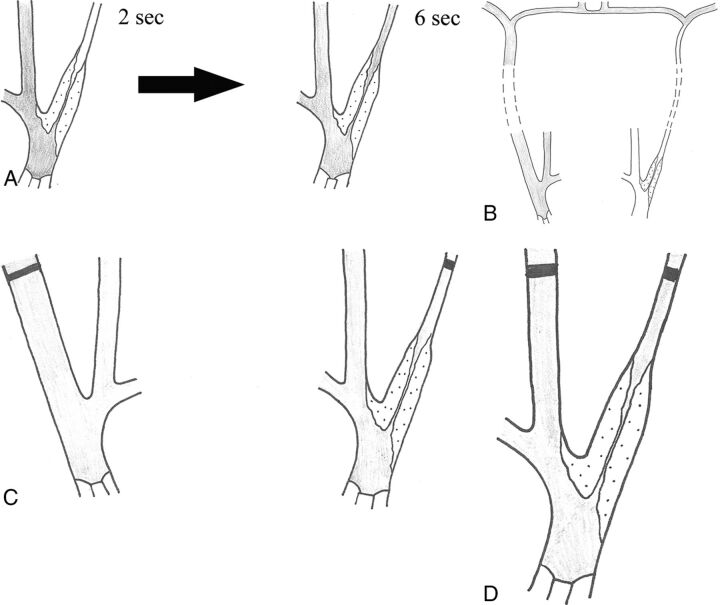
Fig 6.
Schematic drawing of the 4 criteria for near-occlusion on conventional angiography. Delayed filling (A), evidence of intracranial collaterals when the contralateral side is examined (B), ipsilateral distal ICA less than the contralateral distal ICA (C), and ipsilateral distal ICA equal to or less than the ipsilateral ECA (D). In all figures, the contrast is gray.

