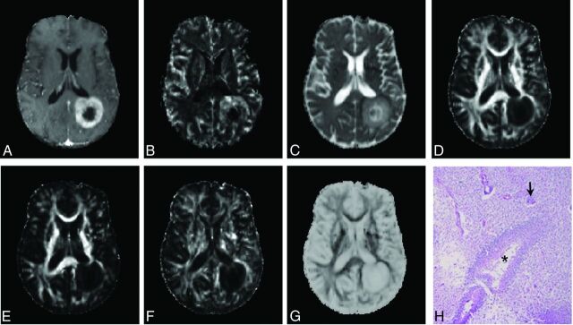
Fig 3.
Axial brain images from a 54-year-old man showing TP. Contrast-enhanced T1-weighted image (A) shows a ring-enhancing lesion in the left parietal lobe. High rCBV (B) and increased MD (C) are observed from the lesion. The enhancing part of the lesion demonstrates decreased FA (D), CL (E), and CP (F) and increased CS (G). Findings in a photomicrograph of a histologic section (H, hematoxylin-eosin stain, 50× magnification) are similar to the patient's de novo glioblastoma, with areas of high tumor cellularity, pseudopalisading necrosis (asterisks), and endothelial proliferation (arrow) and increased mitotic activity.

