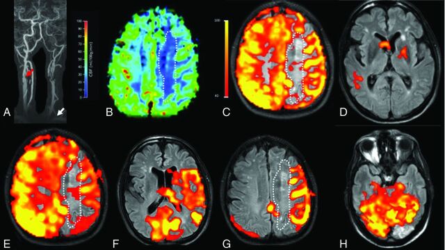
Fig 4.
A 78-year-old male patient with extracranial left ICA occlusion and an extracranial high-grade right ICA stenosis (red arrow) and a left VA stenosis in the V1 segment (white arrow, A). The AcomA and both PcomAs are patent. A perfusion deficit (white dotted line) is seen in the left corona radiata in both the DSC-PWI (B) and the nonselective pCASL map (C). Selective labeling was performed for the left common carotid artery (D), right ICA (E), right VA (F and G), and left VA (H). The intracranial perfusion signal is missing on labeling of the left common carotid artery (D) as a proof of left ICA occlusion. Right ICA labeling (E) shows perfusion of the left anterior cerebral artery and MCA territory. Right VA labeling (F and G) shows recruitment of the posterior circulation for the perfusion of the left MCA territory. The new watershed region with a perfusion deficit and chronic infarcts is seen between the posterior circulation and the right ICA (E and G). A subacute infarct lesion in the left occipital lobe is localized within the perfusion territory of the left VA (H) and can be categorized as a territorial infarct.

