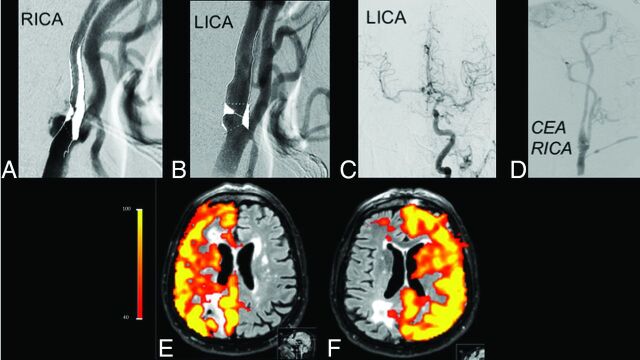
Fig 5.
Initial cDSA shows high-grade (>70%) stenoses of both ICAs (A and B), with flow on the right MCA and anterior cerebral artery from the left ICA (C). After carotid endarterectomy (D), perfusion patterns of the right and left ICAs are normalized (E and F), demonstrating the utility of this noninvasive method for the follow-up of therapy success. RICA indicates right ICA; LICA, left ICA; CEA, carotid endarterectomy.

