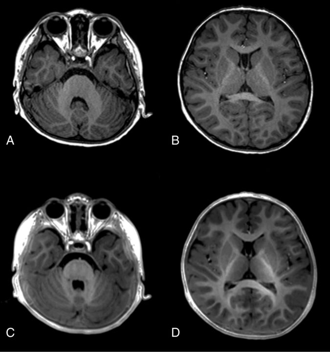
Fig 4.
Images of a 27-month-old boy. MPRAGE (A and B) and PETRA (C and D) images both show prominent hypersignals at all assessed locations, including the cerebellar WM, anterior part of the posterior limb of the internal capsule, genu and splenium of the corpus callosum, and the subcortical WM at the temporal, frontal, and occipital lobes, indicating myelination.

