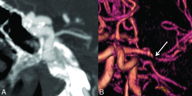
Fig 1.
Imaging findings in Marfan syndrome. A, A 25-year-old man with Marfan syndrome with a 4-mm periophthalmic aneurysm of the right ICA. B, 3D reconstruction of a CTA performed for evaluation of acute ischemic stroke and headache in a patient with Marfan syndrome demonstrates smooth tapering of the right MCA, consistent with an acute dissection (arrow).

