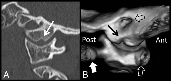
Fig 5.
Sagittal CT (A) demonstrates the medial occipital condyle notch (white arrow) in an 8-year-old boy. A 3D surface-rendered reconstruction image (B) with a medial-to-lateral perspective demonstrates the 3D contour of the occipital condyle notch (black arrow). For orientation, the anterior arch of C1 (white hollow arrow) is to the right and the posterior arch of C1 (white block arrow) is to the left. The internal auditory canal is shown by a hollow black arrow. Note how the notch is wider at its medial aspect compared with the lateral aspect. Ant indicates anterior; post, posterior.

