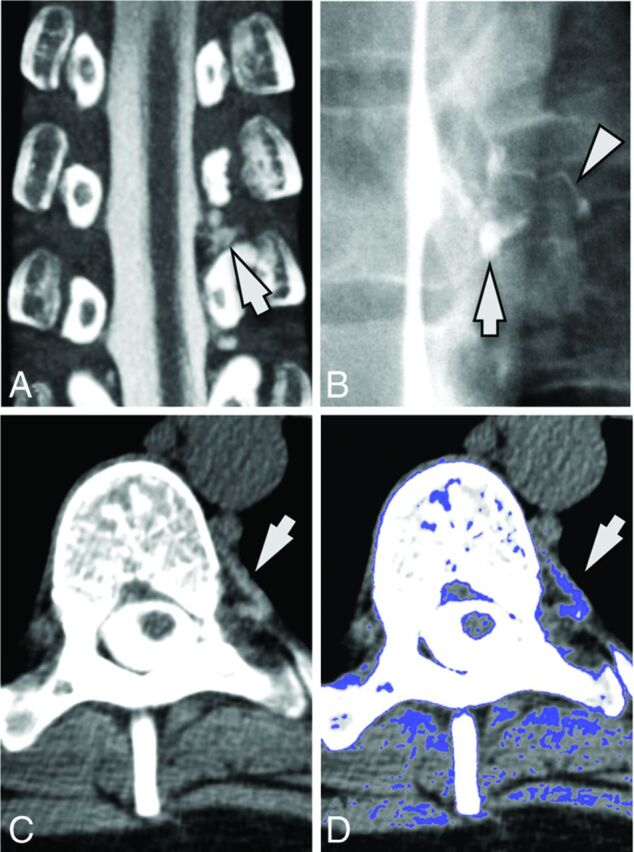
Fig 1.

A 34-year-old woman with SIH. A, Coronal image from a CT myelogram shows a low-flow CSF leak inferior to the left T8 nerve root (arrow). B, A subsequent dynamic myelogram obtained with the patient in the left lateral decubitus position shows the area of the leak (arrow) with a fistula to an adjacent paraspinal vein (arrowhead). C, Axial image from her original CT myelogram reveals a hyperattenuated paraspinal vein (arrow). D, Postprocessed image with thresholded color overlay depicting attenuation values from 60 to 140 HU helps improve the conspicuity of this hyperattenuated vein.
