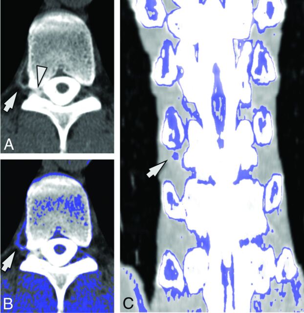
Fig 2.
A 56-year-old man with SIH. A, Axial image from a CT myelogram shows a low-flow CSF leak in the right neural foramen at T10–11 (arrowhead). An adjacent hyperattenuated paraspinal vein (arrow) is seen. B, Postprocessed image with thresholded color overlay depicting attenuation values from 60 to 140 HU improves the conspicuity of this hyperattenuated vein (arrow). C, Coronal image with thresholded color overlay demonstrates that only the vein at the level and side of the fistula (arrow) shows increased attenuation.

