Fig 3.

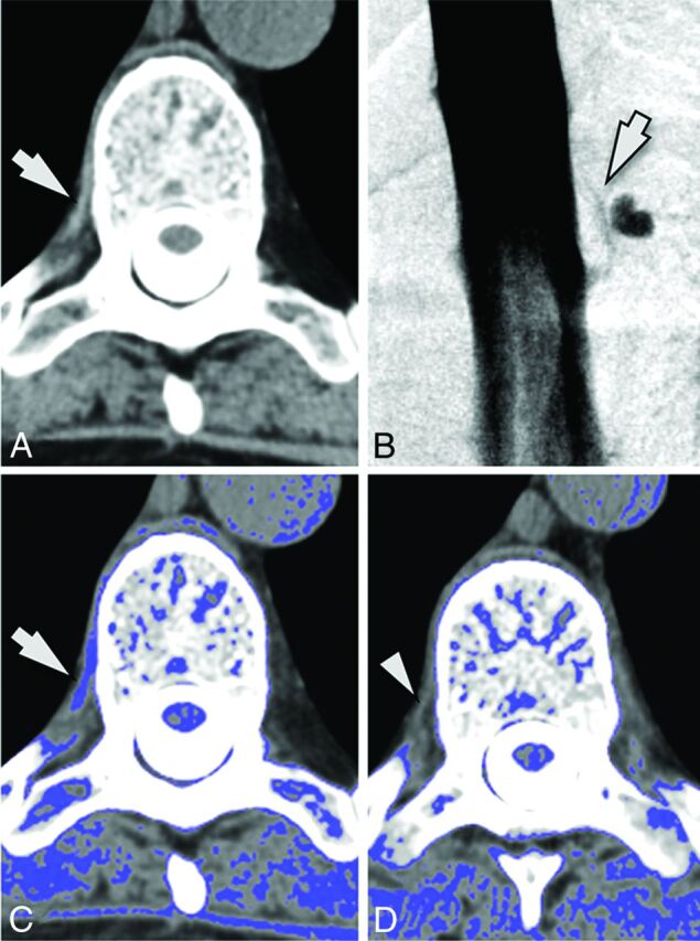
A 59-year-old woman with SIH. A, Axial image from a CT myelogram shows a hyperattenuated paraspinal vein (arrow) at T6–7 on the right. B, A subsequent digital subtraction myelogram shows a CSF-venous fistula at this location (arrow). C, Postprocessed image with thresholded color overlay depicting attenuation values from 60 to 140 HU again helps with the identification of this finding (arrow). D, Axial image from an adjacent level (T8–9) where there was no fistula is provided for comparison. Note that the paraspinal vein is not hyperattenuated (arrowhead) and is not identified on the thresholded color overlay.
