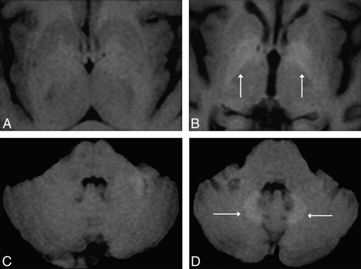
Fig 1.
Axial MR images in a 51-year-old woman with parkinsonism. Unenhanced T1-weighted MR imagings of the first (A and C) and fifth (3 years later; B and D) gadolinium-enhanced MR imagings performed with a nonionic linear GBCA (Omniscan) at the level of the basal ganglia (A and B) and the level of the dentate nuclei of the cerebellum (C and D). The images show progressively increased T1 signal of the globi pallidi and dentate nuclei (white arrows, B and D), undetectable on the first MR imaging.

